Most horse owners have heard horror stories about laminitis—and they should. This devastating hoof condition can transform a sound, athletic horse into a three-legged lame animal in less than 48 hours. The internal structures holding your horse's coffin bone in place literally tear apart, sometimes dropping the bone right through the bottom of the hoof.
But here's what matters: you've got a brief window to stop permanent damage. Miss those early signals? You're looking at months of expensive treatment, possible retirement, or worse.
What Is Laminitis and Why It Threatens Your Horse's Soundness
Picture hundreds of interlocking ridges—like Velcro on a microscopic level—suspending a 1-2 pound bone inside the hoof capsule. That's your horse's laminar connection. These structures, called laminae, work like suspension cables on a bridge. When inflammation attacks them, they stretch, tear, and eventually fail.
The coffin bone, no longer anchored, rotates tip-downward or drops straight through the hoof. Some cases end with bone protruding from the sole—veterinarians call this "penetration," and it's as catastrophic as it sounds.
You'll hear vets distinguish between acute and chronic presentations. Acute means you're in the first 72-hour inflammatory storm. React fast enough, and you might dodge permanent changes. Miss that deadline, and you're managing chronic laminitis—restructured hoof capsules, displaced bones visible on X-rays, and long-term soundness questions.
Among all horse hoof diseases, this one's particularly nasty because it attacks the foundation. A horse can survive many injuries, but ask their feet to bear 800-1,200 pounds without proper internal support? That's asking the impossible.
Prognosis varies wildly. Catch rotation under 5.5 degrees with laminae still mostly intact? You've got decent odds. Hit 11.5 degrees or watch that bone sink straight down? Even top veterinary hospitals struggle with those cases. The amount of displacement matters, but so does your response time and whether you can eliminate whatever triggered the attack in the first place.
One more reality: horses that survive one bout become sitting ducks for round two. Their compromised laminar tissue never quite regains full strength.
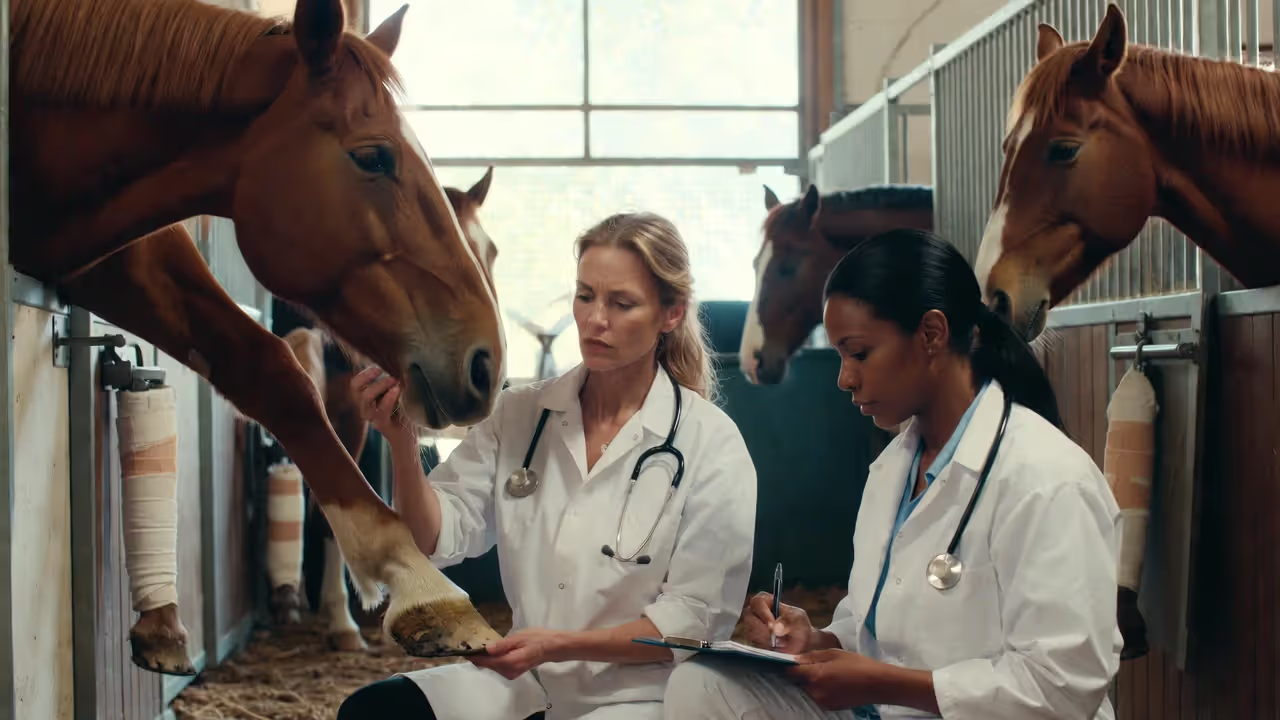
Early Warning Signs Every Horse Owner Must Recognize
Author: Nathan Caldwell;
Source: 3templatedesign.site
Too many owners wait until their horse can barely hobble across the stall. By then, you're playing catch-up with significant damage already done. Learn to spot trouble in the first 12-24 hours instead.
Behavioral Changes That Signal Pain
Watch for the horse that suddenly becomes a statue. Where they'd normally walk over for treats, they now swivel their ears but stay planted. Weight shifting provides another clue—front foot to front foot, never settling into a comfortable rest stance.
Unusual lying-down patterns deserve attention. Your horse spending three hours flat out when they normally stand? Something hurts. Similarly, that previously friendly gelding who now pins his ears when you approach his feet might be telling you they're painful.
Notice whether your horse positions themselves near feed and water or retreats to the stall's back corner. A horse avoiding the ten-foot walk to their hay net isn't being lazy—they're hurting.
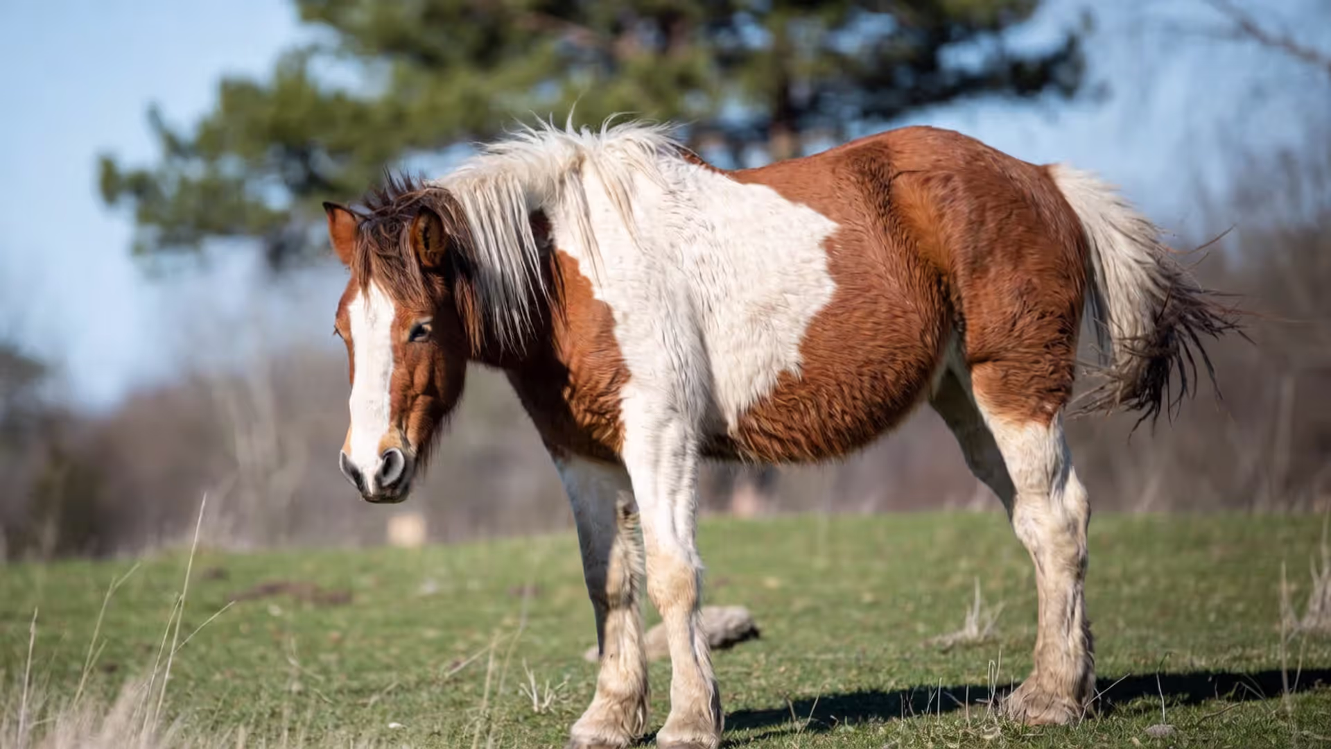
Physical Symptoms in Stance and Movement
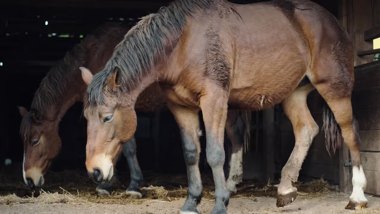
The stereotypical laminitis stance puts front legs stretched forward, hindquarters tucked underneath, creating that "sawhorse" profile. But don't wait for textbook presentation. All four feet can develop laminitis, and hind-limb cases produce different weight distribution.
Gait changes show up before the stance does. Your horse lands toe-first instead of the normal heel-first pattern. They take short, choppy steps like walking on hot coals. Turns become pivots rather than smooth arcs. Hard ground amplifies the lameness; soft footing might disguise it.
Run this test: walk your horse in a tight circle on concrete. Healthy horses execute smooth turns. Laminitic horses look stiff and mechanical, sometimes refusing to turn at all.
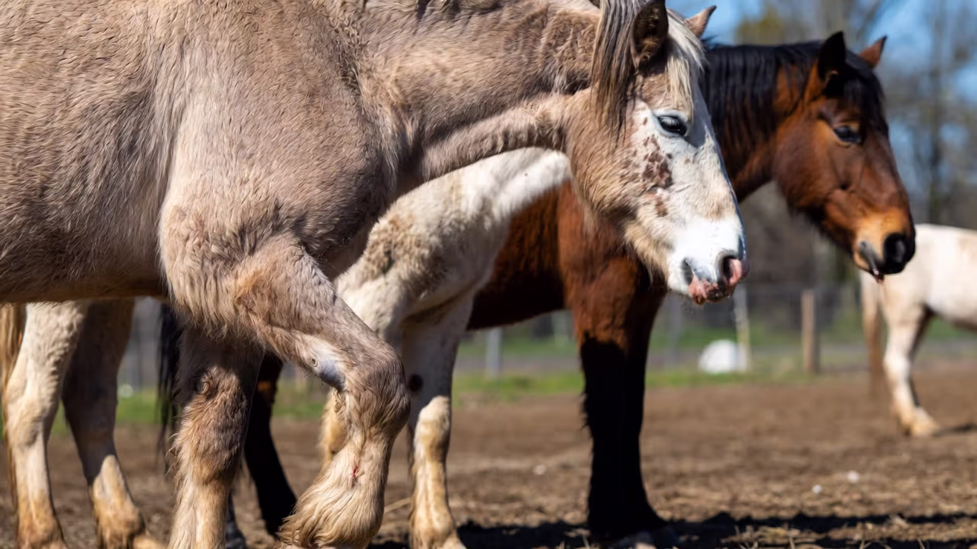
Hoof-Specific Indicators
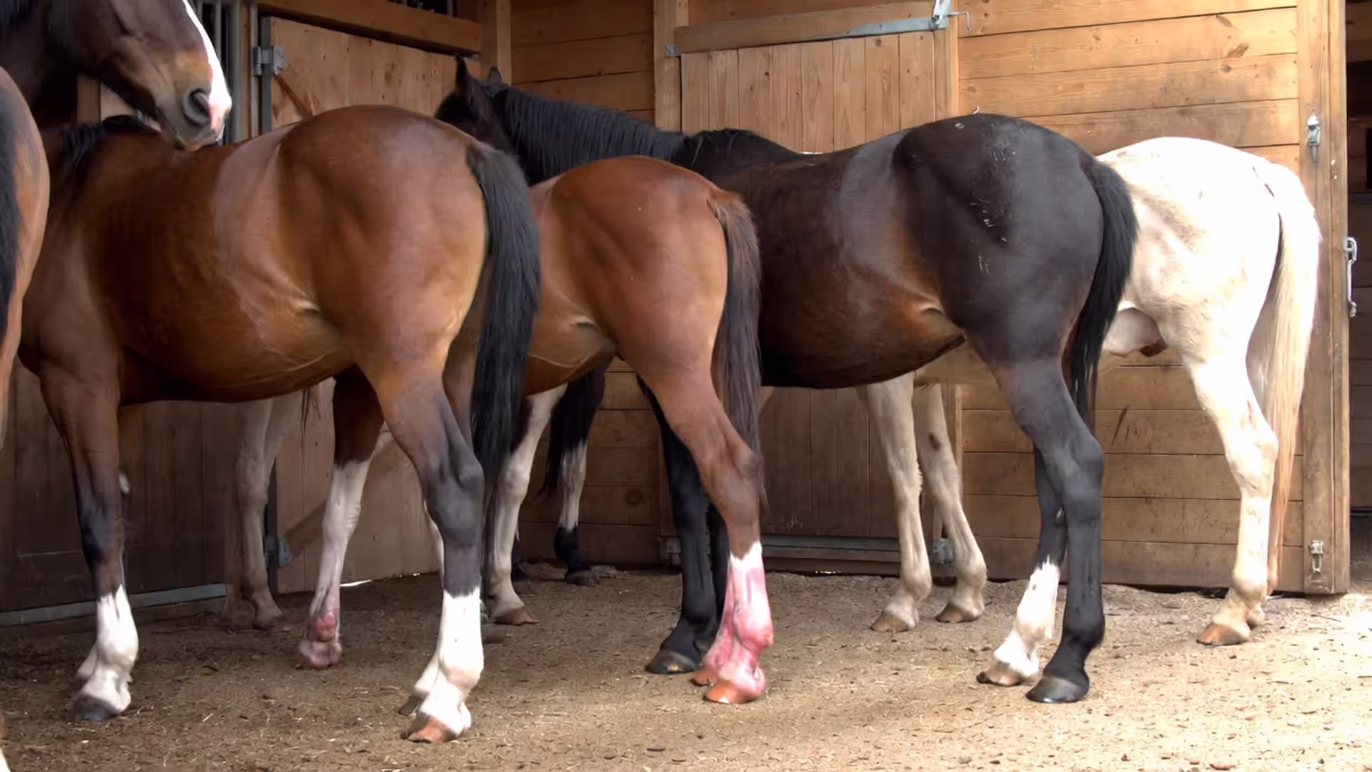
Slide your hand from knee down to coronary band. The hoof wall radiates heat—sometimes enough that you'll feel warmth through winter gloves. Compare front to hind, left to right. One hoof noticeably warmer? That's inflammation.
Find the digital pulse on the pastern's back edge, running alongside the tendons. Normally, you'll barely detect it. During active laminitis, it pounds like a drummer keeping time���strong, bounding, impossible to miss.
Apply thumb pressure to the sole just ahead of the frog's apex. Flinching or foot-snatching indicates sensitivity that shouldn't exist. While you're examining the sole, look for depression or "dishing" along the dorsal (front) hoof wall—evidence of previous rotation that changed growth patterns.
These laminitis symptoms in horses require veterinary evaluation within hours, not days. "Let's see if it's better tomorrow" has ended too many horses' careers.
The Six Most Common Triggers Behind Laminitis Cases
Figuring out what causes laminitis in your specific horse determines whether treatment succeeds or just buys temporary relief. Six major triggers account for most cases, though occasionally multiple factors gang up simultaneously.
Metabolic dysfunction drives roughly 90% of domestic horse laminitis. Equine Metabolic Syndrome (EMS) creates insulin dysregulation—elevated insulin directly damages laminar cells. Pituitary Pars Intermedia Dysfunction (PPID, often called Cushing's disease) similarly disrupts insulin management while causing other systemic problems. These horses develop laminitis from pasture access that wouldn't bother metabolically normal animals. That overweight mare with a neck crest resembling a bodybuilder's trapezius muscle? She's a walking laminitis risk until proven otherwise.
Carbohydrate overload happens two ways: grain bin raids or spring grass binges. Either dumps excessive non-structural carbohydrates into the hindgut. Beneficial bacteria die off, harmful bacteria proliferate, and endotoxins flood the bloodstream. Those toxins trigger the inflammatory cascade attacking laminae. Here's the sneaky part—your horse grazes Monday, appears fine Tuesday, then can't walk Wednesday morning. That 24-48 hour delay between cause and effect confuses owners who insist "nothing changed."
Spring grass deserves special mention. Cool nights below 40°F followed by sunny days cause sugar accumulation in grass blades. Morning grazing (before 10 AM) exposes horses to lower sugar levels than afternoon sessions. April and May destroy more feet than any other months.
Supporting limb laminitis creates a vicious cycle. Fracture the left front, and the right front carries double duty. Exceed the laminar tissue's weight-bearing capacity for days or weeks, and it fails. Now you're treating two crippled legs instead of one. Veterinarians managing severe single-limb injuries live in fear of this complication.
Toxic exposures produce rapid-onset cases. Black walnut shavings—even 5-10% contamination in pine bedding—cause reactions within 8-12 hours of contact. Systemic infections releasing bacterial endotoxins (colitis from Salmonella, retained placentas post-foaling, pneumonia with bloodstream involvement) attack laminae as collateral damage while your horse fights the primary infection.
Corticosteroid administration carries variable risk depending on formulation, dose, route, and individual horse susceptibility. Intra-articular (joint injection) administration poses less danger than intramuscular shots, but metabolically compromised horses occasionally develop laminitis from any corticosteroid exposure. Triamcinolone, dexamethasone, methylprednisolone—all carry some risk. Veterinarians weigh treatment benefits against laminitis danger before prescribing.
Concussion-induced cases require extreme circumstances. Long highway trailer hauls followed by immediate work on hard surfaces might do it. Poorly executed therapeutic shoeing that radically changes weight distribution occasionally triggers inflammation. The old "road founder" diagnosis probably blamed concussion for many metabolically-driven cases we'd now recognize as EMS or PPID.
How Veterinarians Diagnose and Grade Laminitis Severity
Author: Nathan Caldwell;
Source: 3templatedesign.site
Your vet won't diagnose laminitis just from watching a lame horse. Accurate assessment combines hands-on examination, pulse evaluation, and imaging to separate mild from career-ending presentations.
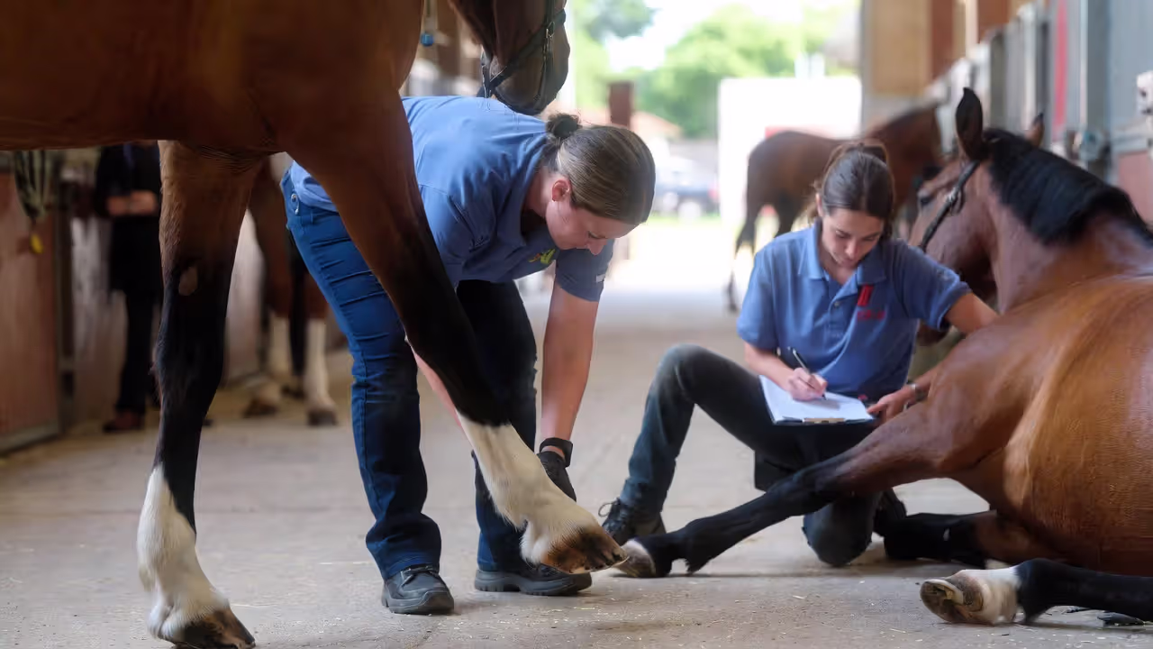
Physical examination starts with observation—stance, gait at walk and trot, turning ability, willingness to move. Hoof testers applied systematically across the sole reveal pain patterns. Increased sensitivity at the toe compared to heels points toward laminar involvement rather than abscesses or bruising.
Digital pulse evaluation happens at the pastern's back edge where arteries run alongside tendons. Normal pulses barely register. Grade 1 laminitis produces easily felt pulses. Severe cases create bounding pulses you can see visually without touching. Pulse strength tracks inflammation intensity and provides real-time treatment response feedback.
Radiographs (X-rays) separate guesswork from facts. Lateral (side) and dorsopalmar (front-back) views reveal coffin bone position relative to the hoof capsule. Veterinarians measure rotation angle, founder distance (the gap between bone and hoof wall at the toe), and sole depth (how much protection exists between bone tip and ground). Serial radiographs taken days or weeks apart document improvement or deterioration.
The Obel system, created back in 1948, categorizes clinical presentation:
Grade
Observable Symptoms
Horse Behavior
Typical Prognosis
Grade 1
Lame at trot, sound at walk; shifts weight frequently between front feet
Moves when asked but steps carefully; lifts feet alternately when standing
Excellent recovery likely with immediate treatment; structural damage usually minimal or absent
Grade 2
Clearly lame at walk with shortened stride; moves without extreme resistance
Reluctant movement; sawhorse stance developing; pain obvious but manageable
Good outcomes if treatment starts within 48 hours; some rotation possible but usually controllable
Grade 3
Severe lameness; moves only with substantial encouragement; resists lifting any foot for fear of bearing full weight on others
Extremely reluctant to take a single step; intense pain response to pressure; may tremble from pain
Guarded prognosis; significant rotation expected; requires months of intensive management even when successful
Grade 4
Refuses all movement; may lie down continuously; cannot support weight on affected feet even briefly
Will not stand voluntarily; shows severe distress if forced upright; may groan or sweat from pain
Poor outlook; complete laminar failure likely; coffin bone may have penetrated sole; humane euthanasia often considered
Veterinarians also factor in time since onset (acute versus chronic), how many feet hurt, and the suspected underlying cause when discussing realistic expectations and treatment plans with owners.
Treatment Protocols: From Emergency Response to Long-Term Management
Effective laminitis treatment options span from immediate first aid to year-long rehabilitation programs. Your response during the first 48 hours often determines whether you're dealing with a minor setback or permanent retirement.
First 48 Hours: Critical Emergency Steps
Before your vet arrives, get the horse onto deep bedding—12+ inches of shavings over rubber mats or a sand-bedded area that cushions every step. This reduces concussive forces on damaged laminae.
Ice therapy (cryotherapy) slows the inflammatory cascade destroying laminar tissue. Fill hoof boots with ice water and keep them on continuously, or stand your horse in a creek. Research from Australia showed 48-72 hours of continuous icing dramatically reduced structural damage when applied early enough. The catch: start within hours of onset, not days.
Pull all grain from the diet immediately. Zero. None. If you suspect pasture triggered this, remove grazing access completely. Offer grass hay—ideally tested below 10% combined sugar and starch, though any grass hay beats continued grazing. Keep water within easy reach so the horse doesn't walk far.
Don't "walk it out." That outdated advice causes additional mechanical trauma to already-failing laminae. Restrict movement to what's absolutely necessary.
Pain Management and Anti-Inflammatory Approaches
Author: Nathan Caldwell;
Source: 3templatedesign.site
Phenylbutazone (bute) and flunixin meglumine (banamine) control pain while reducing inflammation. Veterinarians dose carefully—you want enough relief to prevent abnormal weight-shifting complications, but completely masking pain encourages movement that damages tissue further.
Pentoxifylline improves blood flow through tiny capillaries, theoretically supporting healing in damaged laminae. Aspirin provides mild anti-inflammatory effects and may reduce microclotting in compromised vessels. For chronic cases involving nerve pain, gabapentin addresses neuropathic components NSAIDs don't touch.
DMSO (dimethyl sulfoxide) shows up in acute protocols, applied topically to the coronary band or administered intravenously. It scavenges free radicals and reduces inflammation, though evidence for dramatic benefits remains debated.
Mechanical Support and Therapeutic Shoeing Options
You're trying to support the coffin bone while reducing breakover forces (the effort required to roll the hoof forward during each step).
Deep bedding or glue-on foam pads distribute pressure away from the painful toe region toward the heel and frog. Wooden or Styrofoam frog supports glued directly to the sole help stabilize the coffin bone by loading structures designed to bear weight.
Once acute inflammation settles (typically 7-14 days), farriers apply therapeutic shoeing. Heart bar shoes distribute forces to the frog while providing heel support and reducing load at the toe. Reverse or rolled-toe shoes move breakover backward, reducing tension on the deep digital flexor tendon that pulls the coffin bone's back edge downward into rotation.
Some cases benefit from glue-on shoes that avoid nailing into compromised hoof walls. Polyurethane impression material molded to the sole creates custom support matching that individual hoof's needs.
Trimming focuses on aligning the coffin bone with the hoof capsule using radiographic guidance. Aggressive trimming during acute phases often worsens outcomes; conservative approaches prioritizing comfort generally produce better results. Your farrier works from X-rays, not guesswork, taking small steps every 2-3 weeks rather than making dramatic changes.
Addressing the Underlying Cause
Pain medications and supportive shoeing buy time, but eliminating the trigger determines long-term success. Metabolic horses need dietary overhaul, weight loss protocols, and sometimes pharmaceutical support. Metformin improves insulin sensitivity in some horses. Levothyroxine (thyroid supplementation) aids weight loss in obese cases. PPID horses require pergolide (Prascend) to manage pituitary dysfunction.
Infection-driven cases demand aggressive antimicrobial therapy alongside supportive care for the primary illness. Colitis, pneumonia, or post-foaling complications need resolution before feet improve.
Grain overload cases might benefit from gastric acid suppressants (omeprazole) and hindgut support supplements containing probiotics and yeast cultures.
Bloodwork reveals metabolic dysfunction: resting insulin levels, ACTH testing for PPID, oral sugar tests or insulin tolerance tests that expose insulin dysregulation not visible on basic panels. You can't fix what you haven't diagnosed.
What I emphasize to clients is identifying metabolic risk before symptoms appear. Most laminitis cases are preventable when we recognize susceptible horses early and manage them appropriately. After one episode, that horse requires different care permanently—you're managing predisposition for their remaining years.
— Nicholas Frank, DVM
Frequently Asked Questions About Laminitis
Do horses ever completely recover from this condition?
Recovery depends heavily on severity and intervention timing. Grade 1 cases caught within the first few hours often resolve entirely—the horse returns to previous work levels with no lasting compromise. More advanced cases with measurable coffin bone rotation may achieve "pasture soundness" (comfortable for light riding and turnout) but rarely return to demanding athletic careers. Rotation exceeding 11.5 degrees or sinker presentations (bone dropping straight down) carry poor outlooks even with aggressive treatment. Some survive through intensive lifelong management, but many don't. For chronic presentations, "recovery" means successful ongoing management rather than returning to pre-laminitis condition.
What's the timeline from trigger to obvious lameness?
The inflammatory damage occurs rapidly—within 24-48 hours of the triggering event. However, visible lameness typically lags behind by 24-72 hours, meaning structural damage is already underway when owners first notice problems. That delay explains why "my horse was fine yesterday" accompanies so many emergency calls. Coffin bone displacement progresses over subsequent days to weeks if inflammation continues unchecked. This compressed timeline explains why emergency response matters so critically—you're racing against hours, not days.
Does laminitis always involve too much grass or grain?
Absolutely not. While carbohydrate overload triggers some cases, underlying metabolic disease (EMS and PPID) accounts for the vast majority in domestic horses—these animals develop problems from normal pasture that wouldn't affect metabolically healthy horses. Supporting limb overload, systemic infections, black walnut exposure, and occasionally medications all cause laminitis through completely different mechanisms. The "too much rich grass" explanation oversimplifies a complex disease with multiple pathways to the same endpoint.
How do laminitis and founder differ?
Laminitis technically describes laminar inflammation, while founder refers to mechanical failure—coffin bone displacement resulting from laminar breakdown. In practice, most veterinarians and owners use the terms interchangeably. "Founder" traditionally implied more severe, chronic cases with visible structural changes, whereas "laminitis" might describe milder or earlier presentations. All foundered horses experienced laminitis first, but not every laminitis case progresses to founder when caught and treated early enough.
Laminitis challenges even experienced horsemen, but knowledge converts anxiety into effective action. Recognizing that subtle weight shifts or warm hooves demand immediate veterinary attention—not weekend monitoring—prevents many catastrophic outcomes. Understanding your specific horse's metabolic profile through testing provides individualized management strategies rather than generic internet advice.
Success requires collaboration between owner, veterinarian, and farrier. Owners provide daily observation and implement management modifications. Veterinarians diagnose underlying causes, prescribe evidence-based treatments, and monitor progress through objective measurements. Farriers apply biomechanical principles supporting healing while maintaining functional hoof capsule architecture. Each role carries equal importance—drop one leg of this three-legged stool and the whole effort collapses.
Horses surviving laminitis episodes need permanent heightened vigilance. Seasonal pasture changes, weight fluctuations, or concurrent illnesses can trigger relapses in previously stable cases. This doesn't require living in perpetual fear—it means establishing monitoring systems, maintaining appropriate body condition, and responding quickly to early warnings rather than hoping problems resolve spontaneously.
Prevention remains your most powerful tool. Metabolic testing costs a fraction of laminitis treatment while identifying horses needing special management before crisis strikes. Grazing muzzles seem restrictive until compared against months of painful stall confinement. Weight management feels tedious until you've watched a horse struggle crossing a ten-foot paddock.
Horses most likely to avoid laminitis belong to owners who test hay, honestly assess body condition scores, adjust management seasonally, and maintain veterinary relationships before emergencies occur. These practices don't guarantee immunity—some cases strike despite excellent care—but they shift odds substantially toward your horse's favor.
Colic remains the number one medical cause of death in horses, accounting for nearly 30% of equine fatalities. Early recognition and prompt treatment dramatically improve outcomes. Learn the warning signs, common causes, emergency response protocols, and 12 proven prevention strategies every horse owner needs.
Content on 3templatedesign.site is provided for general informational and educational purposes only. The information on this website may include topics related to horse care, feeding, grooming, horse breeds, training methods, riding techniques, and common horse health conditions.
This content is not intended to be veterinary, medical, training, or professional animal care advice. Horse ownership and riding involve responsibilities and potential risks for both riders and animals.
Users are responsible for evaluating their own experience, equipment, and the condition of their horse before applying any information from this website. Use of this website does not create any professional, veterinary, or advisory relationship with trialstribulations.net.
We are not responsible for any injuries, damages, or losses resulting from the use of information provided on this website. Horse owners and riders are encouraged to consult qualified veterinarians, trainers, or equine professionals when making decisions about horse care or training.